Sacroiliac Luxation Repair
Rollins, Amanda et al. “Evaluation of Fluoroscopic-Guided Closed Reduction versus Open Reduction of Sacroiliac Fracture-Luxations Stabilized with a Lag Screw.” Veterinary and comparative orthopaedics and traumatology : V.C.O.T vol. 32,6 (2019): 467-474. doi:10.1055/s-0039-1693471
Key Point: Fluoroscopic-guided sacroiliac luxation repair leads to consistently more optimal screw placement, as well as a lower occurrence of lag screw loosening as compared to traditional approaches.
Sacroiliac (SI) fracture-luxations are a fracture between the sacrum (pelvic vertebral column) and the ilium (pelvis) – see image below. Most of these injuries occur after a dog is hit by a car, and can happen concurrently with other trauma to the body (lung injury, cardiac injury, bleeding). SI luxations can be associated with injury to the sacral spinal nerve roots and lumbosacral spinal trunk. Surgery is indicated for dogs with signs of severe pain or displacement that either compromises pelvic canal diameter or hip joint alignment.
The approach clinically to a SI luxation is reduction of the fracture back to the original anatomy, and fixation with a screw and washer. Many variations exist in the approach, but previous mechanical studies have shown that a lag screw of the largest diameter should be inserted to a depth of at least 60% of the sacral body width. The target for optimal screw placement in the sacrum in a about 1cm2 in a average large sized dog – not much room for error! Missing this target zone can leads to iatrogenic (inadvertently done) trauma to the nerve roots or not achieved adequate fixation, which leads to loosening and potential surgical failure.
Where available, intraoperative fluoroscopy (intra-operative x-ray) has been used with great benefit for reduction and stabilization of sacroiliac separation. This helps us hit our target. Minimally invasive techniques, through keyhole incisions, avoid the need for large incisions and provide the ability to guide the placement of the screw with real-time imaging. This removes most of the guesswork involved in driving the screw deep into the sacral body, and eliminates an extensive surgery.
While a direct comparison has been previously made in cadaver studies, no clinical study has directly compared open versus closed reduction until now. Dr. Rollins and Dr. Balfour in 2019 published a paper where they compared the traditional approach to the imaging guided approach through a keyhole incision. Fluoroscopic-guided approaches lead to consistently more optimal screw placement, as well as a lower incidence of screw loosening (8% in vs 53%). This mimics what we see clinically and highlights an optimal and consistent surgical outcome with this technique. Ultimately, surgeries with optimal outcomes and patient safety are what we strive for. At Arizona Canine Orthopedics we utilize intra-operative x-ray in multiple surgeries, to make every surgery as safe as possible!

Image credit: Dr. Petrovsky.
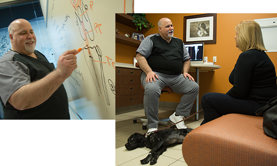
Brian Petrovsky, DVM
Practice Limited to Surgery
Get Informed
Schedule a Consultation
Make no mistake, all surgical procedures are serious. Get the information you need and know your options. Then make an informed decision. Like any service, not all veterinary services are equal. Call to schedule or ask questions.
Call our clinic at 480.998.5999
or Email us.
FAQ
What Makes ACOSM Stand Out?
We pride ourselves on doing things differently. We insist on providing premier service to our patients and their caregivers. There's a saying, "Price is what you pay; value is what you get." At ACOSM, delivering on value is our mission.
We're proud of our experience, skill, and outcomes; it puts us in a category of one, which means you'll experience things with us not promised by any other veterinarian in AZ.
Dog Blog
Distal Femoral Osteotomy for Comprehensive Treatment of Medial Patella Luxation
Three-Dimensional Printed Patient Specific Guides for Acute Correction of Deformities
Comparison of Arthroscopy and Arthrotomy for Diagnosis of Medial Meniscal Pathology
Second Look Arthroscopic Findings After Tibial Plateau Leveling Osteotomy
Resources
New Patient Registration Forms
- New Patient Packet

- New Patient Packet - Online
 New online form!
 - Patient Referral Form
- Patient Referral Form - Online
Financing Options
- Home
- About Us
- Our Commitment
- Common Procedures & Conditions
- WatchDog™
- 3D Planning and Modeling
- FAQ
- Blog
- Contact Us
Copyright © 2025 Arizona Canine Orthopedic Sports Medicine (ACOSM). All rights reserved. Sitemap