Map
Canine elbow dysplasia is a hereditary joint disease affecting young dogs during growth and development. Eighty percent of affected dogs have bilateral disease (both elbows).
The elbow joint consists of three bones - the radius, ulna and the humerus. Growth abnormalities may cause imperfections in the way the bones fit together, and the incongruency results in abnormal pressure within the joint that may either wear down cartilage or result in bone/cartilage fragmentation.
Over time, elbow dysplasia causes inflammation, pain, lameness, and osteoarthritis.
Dogs with elbow dysplasia have a limp and often an accompanying head bob. As they plant the paw of the healthy (or less painful) front leg on the ground, their head bobs downward - “down is sound.” as the expression goes. As the dog puts the paw of the painful leg on the ground, the head and neck lift upward. Head bobbing is easier to spot when the dog walks or trots in a straight line and becomes more challenging to see when they run. Dogs with dysplastic elbows are reluctant to walk or play for long periods.
Breeds frequently affected include:
- German Shepherds
- Rottweiler
- Bernese Mountain Dogs
- Golden Retrievers
- Labrador retrievers
- Giant breed dogs
While elbow dysplasia sounds like one disease, it consists of several associated pathologies including:
- Osteochondritis dissecans (OCD)
- Ununited anconeal process (UAP)
- Fragmented medial coronoid process (FMCP)
Diagnosis
- Gait Evaluation - the doctor observes the mechanics of how your dog walks, noting any abnormalities.
- Physical & Orthopedic Exam - the surgeon performs specific tests to identify and localize the cause of lameness and pain and checks the range of motion in flexion (bending) and extension (straightening) of the elbow.
- Radiographs - X-rays show the overall health of the elbow joint and the degree of pre-existing osteoarthritis. Incongruency is often underestimated and a distinct FMCP may not be seen on x-ray. What is seen are secondary joint remodeling changes that are consistent with elbow dysplasia.
- CT Scan - joint incongruency and FMCP are best seen non-invasively by CT. CT is more sensitive and offers numerous advantages compared to standard radiographs.
- Arthroscopy - Arthroscopy is used both as a diagnostic tool and to treat elbow dysplasia. The surgeon makes small incisions on the inside of the elbow and inserts the arthroscope, which provides high resolution, magnified and brightly illuminated images of the inside of the joint. The doctor can then remove loose fragments of bone and cartilage, and joint surfaces can be debrided (cleaned-up).
Canine osteochondritis dissecans (OCD)
Osteochondritis Dissecans (OCD) is a juvenile orthopedic disease that may affect several joints. In young, skeletally immature dogs, the ends of the bones grow to length and normally replace specific areas of cartilage. When the process fails, cartilage thickens but never forms bone. The deepest layer is poorly-nourished, and that's because cartilage gets the nutrients it needs to stay viable (alive) by diffusion from the joint fluid. Because the cartilage is so thick and poorly nourished, its deepest layer dies, developing cracks that migrate to the joint surface. The uppermost cartilaginous layer loses its connection to the underlying bone and forms a flap that may partially or completely detach into the joint. The condition causes pain, inflammation, and lameness.
Canine ununited anconeal process (UAP)
Ununited anconeal process (UAP) is an elbow disorder where the growth plate between the upper part of the ulnar notch within the elbow joint (called the anconeal process) fails to close normally, which results in joint instability, inflammation, and pain.
Fragmented medial coronoid process (FCP)
The ulna bone has two small protrusions called coronoid processes. In FCP, a coronoid process develops a crack and separates from the rest of the bone. When it occurs on the medial (toward the dog's midline) coronoid process, surgeons diagnose the condition as a fragmented medial coronoid process (FMCP). Incongruency and excessive pressure on the innermost part of the joint causes erosion.
Prognosis
Elbow dysplasia is a genetic orthopedic condition. In other words, a dog who’s been diagnosed with elbow dysplasia always has elbow dysplasia.
There is no cure for this heritable, conformational problem.
There are, however, a wide variety of medical and surgical treatment options. Overall, most dogs with elbow problems lead a reasonably normal life, but every form of elbow dysplasia may cause osteoarthritis.
Treatment options
Treatment options for canine elbow dysplasia depend on the actual definitive diagnosis, which is why a correct diagnosis, preferably by an experienced surgeon, is crucial.
Non-surgical options are not curative because they don't correct the underlying problem/s within the joint. Non-surgical treatments include:
- Physical therapy
- Weight management
- Exercise modification
- Glucocorticoid steroid injections
- Veterinary prescribed medication/s
- Supplements such as glucosamine
- PRP, hyaluronic acid, or stem cell injections
Surgical treatment options
- Arthroscopy
- Osteotomy of the ulna such as the bi-oblique dynamic proximal ulna osteotomy (BODPUO) and proximal abducting ulna osteotomy (PAUL) or of the humerus (sliding humeral osteotomy).
- Arthroscopy and osteotomy combination
Surgery can remove unstable and/or displaced bone-cartilage fragments that inflame the joint while other procedures improve bone alignment or remove/reattach parts of the bone that irritate.
Complications
Complications exist with every surgical procedure and may involve anesthesia, the process itself, or something occurring during recovery. Several surgical methods are available to treat canine elbow dysplasia, so be sure to discuss possible associated complications with the surgeon.
Recovery
Regardless of which treatment you choose, weight management is essential for dogs with elbow dysplasia. Weight loss decreases stress placed on diseased joints. Ask your veterinarian about safe and healthy ways to help your dog lose weight.
You'll need to restrict activity following surgery, and the surgeon will instruct you to increase activity as your pet recovers. Controlled, slow-paced, and short-duration leash walks will be one of the first things you'll do to help your pet improve muscle strength and range of motion.
Ask the surgeon whether physical therapy will help your dog because PT may benefit your dog’s recovery. Some forms include:
- Massage
- Cold laser therapy
- Underwater treadmill
- Ice or heat treatments
- Passive range of motion (PROM) stretches
- Balance, strengthening, and proprioception exercises
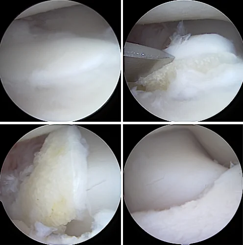
This is an example of a minimally displaced - or in situ - fragmented medial coronoid process (FCP) of the ulna, top left. With minor probing and levering (top right), the unstable FCP is easily dislodged. The FCP is then mobilized and removed by traction with shaver contouring-reduction of the ulna cornoid (bottom images).
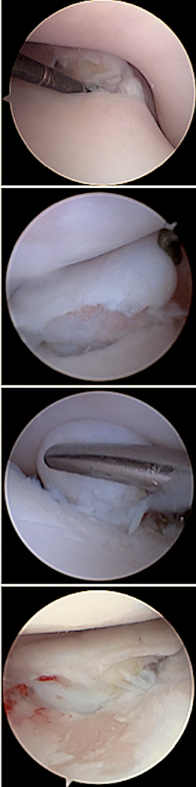
Fragmented medial coronoid processes (FCP) of the ulna are a common finding in dogs with Elbow Dysplasia. FCPs may present very differently in individual dogs - as seen here - and range from minimally displaced incisural fragments (top), to very large and displaced apical bone-cartilage fragments (middle) and extensive articular cartilage wear known as Medial Compartment Disease (bottom).
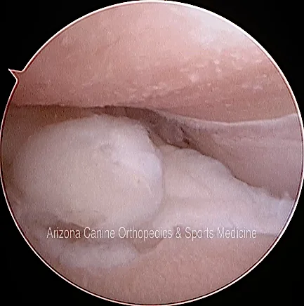
Severe Medial Compartment Disease with extensive loss of articular cartilage can be a consequence of Elbow Dysplasia, joint incongruency and medial coronoid process disease.
Get Informed
Schedule a Consultation
Make no mistake, all surgical procedures are serious. Get the information you need and know your options. Then make an informed decision. Like any service, not all veterinary services are equal. Call to schedule or ask questions.