Map
What does it do?
The knee - or stifle - joint of the dog relies upon soft tissue structures for stability and integrity. Along with the CrCL, other major soft tissue structures stabilizing the stifle are the caudal cruciate ligament and the meniscal cartilages, all of which are intra-articular (within the the joint), and the medial and lateral collateral ligaments (external to the joint). The quadriceps, hamstrings and calf muscles also play a significant role in the forces acting on the joint.
What does it look like?
The CrCL is composed of two bands - a craniomedial and a caudolateral band. The ligament functions to stop cranial (anterior) movement of the tibia relative to the femur, it limits hyperextension of the joint and controls internal rotation of the tibia. The CrCL is thought to be able to resist a force more than four times the weight of the dog before it ruptures.
The CrCL rupture may present in several different ways. There may be a single incident which causes a sudden, complete rupture of the ligament or a complete tearing may occur more slowly over time. Dogs may also partially tear or stretch the ligament due to trauma. With a partial CrCL tear, the dog typically experiences intermittent lameness only. The majority of partial ruptures will progress, if untreated, to complete tears within a matter of weeks to months.
Obesity, conformational, hormonal and genetic factors increase the risk of a CrCL tear.
Meniscal Cartilages
The crescent-shaped, medial and lateral meniscal cartlages are also key intra-articular structures that contribute to joint stability and help to evenly distribute weight bearing loads across the surfaces of the tibia.
The menisci are wedge-shaped and are both thicker at the outer edges and posteriorly. The meniscal cartilages have nerve fibers that tell the brain how load is transmitted through the joint. The medial meniscus is often secondarily damaged following CrCL tearing and affects as many as 83% of dogs. The medial meniscus may be at greater risk because it is firmly attached to the tibia and may become crushed during the abnormal caudal femoral subluxation or displacement.
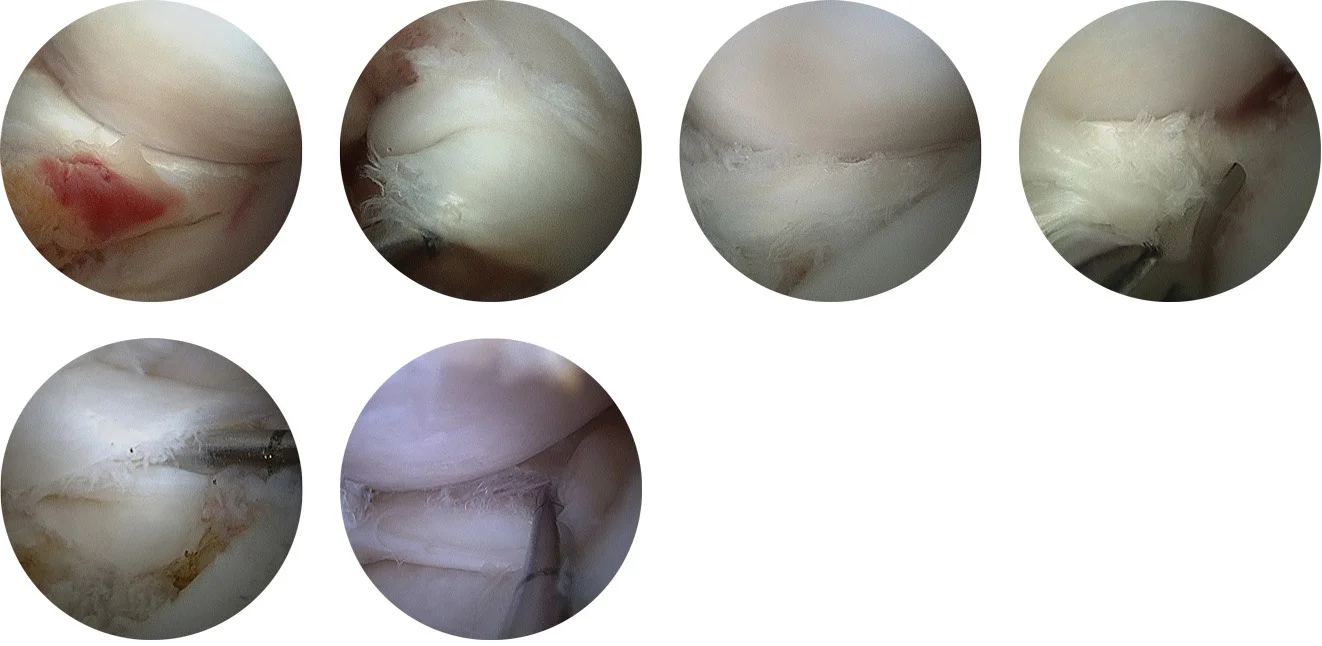
Both the normal schematic and actual appearance as seen by arthroscopy of the meniscal cartilages (left) and typical meniscal tear configurations (right) are depicted schematically below.
Healthy
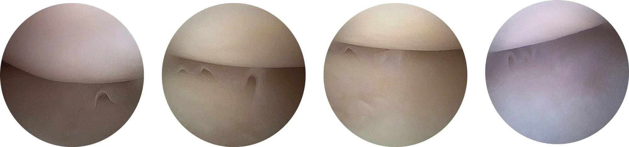
Torn

Do your research.
Then decide.
We’ll help you understand exactly what is going on with your pet. If you’d like to do your own research, read on. Learn about your dog’s anatomy and how we identify exactly what is wrong and all options for treatment.
We strive for full transparency and to provide all of the information you need to make an educated decision. Note: this information gets pretty technical. Have a question? Let us know. Click here.
Get Informed
Schedule a Consultation
Make no mistake, all surgical procedures are serious. Get the information you need and know your options. Then make an informed decision. Like any service, not all veterinary services are equal. Call to schedule or ask questions.